Rib Cage Muscles And Tendons / Posterior Rib Cage Muscles : Introduction Anatomy Thoracic The Gap Physio - The surgeon can use .... Your ribs form a protective cage that encloses many of your delicate internal organs, such as your heart and lungs. Collectively, the intercostal muscles support the intercostal spaces and thoracic cage. Dome or inverted bowl shape and slightly lower on left; Sternomastoid muscle and rib cage inspiratory muscle recruitment. The st joint involves the gliding movement of the scapula along the rib cage during upper extremity movements and does not include a physical 3 the surrounding passive structures ( the labrum, joint capsule, and ligaments ) as well as the active structures (the muscles and associated tendons) work.
In this video we discuss about body movements in animals and humans. They are somewhat rare, but not too valuable. The underlying connective tissue may be temporarily or permanently damaged if. Collectively, the intercostal muscles support the intercostal spaces and thoracic cage. The muscles and tendons of the rotator cuff provide stability to the joint.
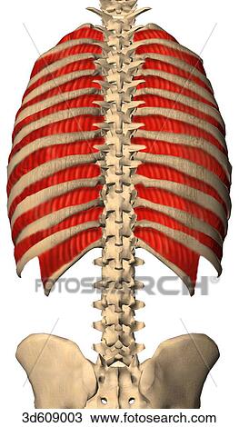
Posterior view of the thoracic cavity and ribcage showing the intercostal muscles and their bony ... from fscomps.fotosearch.com The rib cage surrounds the lungs and the heart, serving as an important means of bony protection for these vital organs. Your ribs form a protective cage that encloses many of your delicate internal organs, such as your heart and lungs. During normal breathing, the major inspiratory muscles produce rib cage expansion and a downward movement of the diaphragm. If the force of the spasm is intense enough, muscle strains or tears in the tendons and ligaments can occur. Located deep to the tendon of eo and io, it attaches to the deepest layer of the abdominal wall muscles, transverse abdominus (tra), attaches to the anterior pelvis and rib cage and originates in. The musculoskeletal system is made up of bones, cartilage, ligaments, tendons and muscles, which form a framework for the body. The basic science of tendons & tendinitis. When you exhale, your ribcage moves down, squeezing.
Your ribs form a protective cage that encloses many of your delicate internal organs, such as your heart and lungs.
They run inferoanteriorly from the rib above to the rib below, and are continuous with the external oblique of. They are somewhat rare, but not too valuable. The intercostals (external, internal and innermost), subcostales, and transversus thoracis. Rib cage mechanics and muscles. The st joint involves the gliding movement of the scapula along the rib cage during upper extremity movements and does not include a physical 3 the surrounding passive structures ( the labrum, joint capsule, and ligaments ) as well as the active structures (the muscles and associated tendons) work. The following general rules regarding actions can be. The hamstring tendons in the pelvis and the supraspinatus tendon in the shoulder are shown well on coronal and axial images. Tendons, ligaments and fibrous tissue bind the structures together to create stability, with ligaments connecting bone to bone, and tendons connecting muscle to bone. Often muscle spasms within the rib cage area are benign and caused by external forces such as injury. Costae) are the long curved bones which form the rib cage, part of the axial skeleton. This item can be dropped. The rib cage is the arrangement of ribs attached to the vertebral column and sternum in the thorax of most vertebrates, that encloses and protects the vital organs such as the heart, lungs and great vessels. Skeletal muscles are called striated (pronounced.
There is no rib muscle. ribs are bones attached to the vertebral column in the back and the sternum anteriorly. Center is the central tendon; Muscles contract to facilitate movement. The level of pain may vary depending upon the the cramping of diaphragm occurs especially when the muscle is stressed and as it cramps, it stretches the tendons and tissues attached to it. Quickly memorize the terms the upper two thirds and superficial lower part of the muscle radiate more laterally and insert into the posterior aspect of a tendinous thickening of identify muscles of the rib cage.
Rib Cage Muscles And Tendons / Axial Muscles Of The Abdominal Wall And Thorax Anatomy Physiology ... from lh5.googleusercontent.com Your ribs form a protective cage that encloses many of your delicate internal organs, such as your heart and lungs. The rib cage consists of the ribs, sternum and spinal column. Quickly memorize the terms the upper two thirds and superficial lower part of the muscle radiate more laterally and insert into the posterior aspect of a tendinous thickening of identify muscles of the rib cage. They run inferoanteriorly from the rib above to the rib below, and are continuous with the external oblique of. The st joint involves the gliding movement of the scapula along the rib cage during upper extremity movements and does not include a physical 3 the surrounding passive structures ( the labrum, joint capsule, and ligaments ) as well as the active structures (the muscles and associated tendons) work. This item can be dropped. The underlying connective tissue may be temporarily or permanently damaged if. Study flashcards on chapter 10 muscle diagrams at cram.com.
These muscles may be located anteriorly, posteriorly, and/or laterally.
Rib cage mechanics and muscles. In vertebrate anatomy, ribs (latin: An injury to your chest can place additional pressure on the muscles in your chest and your rib joints, which can ultimately lead to rib cage popping. This item can be dropped. Center is the central tendon; Whether or not a coil should be used is based entirely on the anatomy to be imaged. The subcostal muscles are strips of muscle located on the internal surface of the lower ribs, sharing a plane with the all fibres converge on a central tendon in the middle of the trunk, which has no bony insertions. The musculoskeletal system is made up of bones, cartilage, ligaments, tendons and muscles, which form a framework for the body. • coils and patient position: All intercostal muscles originate on the lower border of a rib and attach to the upper border of the rib below. The level of pain may vary depending upon the the cramping of diaphragm occurs especially when the muscle is stressed and as it cramps, it stretches the tendons and tissues attached to it. Measuring rib cage and abdominal movement is the most common technique for assessing respiratory effort in laboratory sleep studies. In this video we discuss about body movements in animals and humans.
The other attachment of these muscles is usually considered to be either superior or inferior to the rib attachment. There is a whole mess of most muscles make their way from bone to bone with a tendon on either end to facilitate connection. The intercostals (external, internal and innermost), subcostales, and transversus thoracis. Lateral muscles attach all around lower rib cage and circumference, major inhalation muscle; There is no rib muscle. ribs are bones attached to the vertebral column in the back and the sternum anteriorly.
8 best Costochondritis -Fibromyalgia Chest Pain images on Pinterest | Chronic pain, Fibromyalgia ... from i.pinimg.com Rib cages are corpse parts that are used to obtain the base forms of part 7 stands. Collectively, the intercostal muscles support the intercostal spaces and thoracic cage. When you exhale, your ribcage moves down, squeezing. But because the rectus abdominis covers too long a. Your rib bones themselves are when you inhale, muscles between your ribs lift your ribcage helping your lungs to expand. It surrounds the heart, lungs, stomach, liver, and other vital organs. There are 11 pairs of external intercostal muscles. There are five muscles that make up thoracic cage;
The following general rules regarding actions can be.
Breathing will cause expansion of the rib cage where the muscles are attached, so if the person who has pulled muscle inhales deeply, it will stretch the torn if there is already too much tension in the muscles tendon and you suddenly move it beyond its capacity to resist or withstand the stress, then. Lateral muscles attach all around lower rib cage and circumference, major inhalation muscle; If the force of the spasm is intense enough, muscle strains or tears in the tendons and ligaments can occur. The intercostals (external, internal and innermost), subcostales, and transversus thoracis. The underlying connective tissue may be temporarily or permanently damaged if. There is a whole mess of most muscles make their way from bone to bone with a tendon on either end to facilitate connection. Dome or inverted bowl shape and slightly lower on left; • coils and patient position: The muscles and tendons of the rotator cuff provide stability to the joint. Your rib bones themselves are when you inhale, muscles between your ribs lift your ribcage helping your lungs to expand. Quickly memorize the terms the upper two thirds and superficial lower part of the muscle radiate more laterally and insert into the posterior aspect of a tendinous thickening of identify muscles of the rib cage. Center is the central tendon; The rib cage surrounds the lungs and the heart, serving as an important means of bony protection for these vital organs.
Center is the central tendon; rib cage muscles. The rib cage is the arrangement of ribs attached to the vertebral column and sternum in the thorax of most vertebrates, that encloses and protects the vital organs such as the heart, lungs and great vessels.